A New Blueprint for Turning an Orphan Brain Receptor into a Blockbuster Drug Target

Forthcoming in Drug Discovery Today: A New Blueprint for Turning an Orphan Brain Receptor into a Blockbuster Drug Target

I am pleased to announce that my article, “Accelerating the Orphan GPCR Pipeline: GPR149 as a Case Study in Dual-Domain Target Validation,” has been accepted for publication in Drug Discovery Today, a leading journal for pharmaceutical strategy and drug development. The article presents a comprehensive, industrial-strength framework for de-orphanizing GPR149—a little-understood receptor that has remained a biological mystery for over two decades—and transforming it into a high-value therapeutic asset with blockbuster potential.

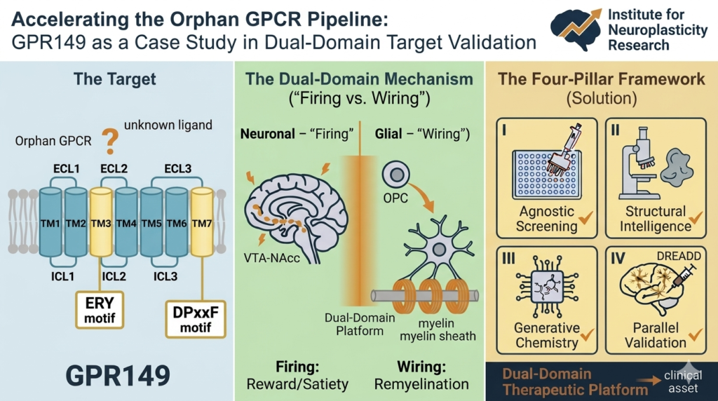
GPR149 is strategically expressed in three critical areas of the brain. First, it is found in the hypothalamus, the body’s master regulator of energy balance, where it appears to influence appetite, satiety, and metabolic set-points. Second, it is concentrated in the ventral tegmental area (VTA) and nucleus accumbens (NAcc)—the brain’s primary reward and motivation circuitry—where it may modulate cravings, reward-seeking behavior, and the compulsive drive that underlies addiction. Third, GPR149 is present in oligodendrocyte precursor cells (OPCs), the glial cells responsible for repairing the brain’s white matter through a process called remyelination.

This unique expression pattern points to a receptor that sits at the intersection of metabolism, motivation, and neural repair. A drug designed to modulate GPR149 could potentially address multiple high-value indications simultaneously. By targeting GPR149 in the hypothalamus and reward circuits, such a drug might reduce stress-induced emotional eating—a key driver of obesity—while also dampening cravings for addictive substances. By acting on OPCs, the same drug could promote remyelination, offering a therapeutic avenue for multiple sclerosis and other demyelinating diseases. This “dual-domain” profile—addressing both the synaptic “firing” of reward circuitry and the structural “wiring” of white matter—distinguishes GPR149 from conventional metabolic and CNS targets.

The article outlines a practical, parallelized Four-Pillar Framework to systematically de-risk GPR149, combining high-throughput screening, cryo-electron microscopy, AI-driven generative chemistry, and circuit-level behavioral validation. The framework is designed to compress discovery timelines, front-load critical failure points, and deliver value even when a clinical candidate remains elusive. Importantly, the article also addresses the receptor’s expression in the pituitary and ovaries, identifying potential on-target fertility effects that can be managed through blood-brain barrier restriction or tissue-specific bias.

The forthcoming publication represents a strategic roadmap for pharmaceutical R&D teams seeking to unlock the therapeutic potential of the “dark GPCRome.” I look forward to sharing the full article when it appears online. For those interested in discussing partnership opportunities, licensing, or collaborative development of GPR149-targeted therapeutics, please contact me directly.

Michael A. S. Guth, Ph.D., J.D.
mike[no spam]@michaelguth.com

𝐅𝐨𝐜𝐮𝐬 𝐨𝐧 𝐄𝐦𝐩𝐨𝐰𝐞𝐫𝐦𝐞𝐧𝐭 & 𝐄𝐝𝐮𝐜𝐚𝐭𝐢𝐨𝐧: 𝐀𝐫𝐞 𝐲𝐨𝐮 𝐛𝐮𝐢𝐥𝐝𝐢𝐧𝐠 𝐲𝐨𝐮𝐫 𝐛𝐫𝐚𝐢𝐧 𝐮𝐩, 𝐨𝐫 𝐚𝐫𝐞 𝐲𝐨𝐮 𝐥𝐞𝐭𝐭𝐢𝐧𝐠 𝐢𝐭 𝐛𝐫𝐞𝐚𝐤 𝐝𝐨𝐰𝐧?

𝐅𝐨𝐜𝐮𝐬 𝐨𝐧 𝐄𝐦𝐩𝐨𝐰𝐞𝐫𝐦𝐞𝐧𝐭 & 𝐄𝐝𝐮𝐜𝐚𝐭𝐢𝐨𝐧: 𝐀𝐫𝐞 𝐲𝐨𝐮 𝐛𝐮𝐢𝐥𝐝𝐢𝐧𝐠 𝐲𝐨𝐮𝐫 𝐛𝐫𝐚𝐢𝐧 𝐮𝐩, 𝐨𝐫 𝐚𝐫𝐞 𝐲𝐨𝐮 𝐥𝐞𝐭𝐭𝐢𝐧𝐠 𝐢𝐭 𝐛𝐫𝐞𝐚𝐤 𝐝𝐨𝐰𝐧? The concept of Cognitive Reserve is the single most empowering framework for understanding how we age.

“Cognitive Reserve” is the buffer your brain builds—a dynamic network of strong neural connections that protects your ability to think, remember, and solve problems, even if your brain begins to show signs of age or damage.
We are not powerless. Your brain possesses Neuroplasticity, an incredible lifelong ability to adapt and rewire. It is actively changing right now, based on your lifestyle choices, experiences, and thoughts. This change can be positive, or it can be negative.

Think of it like a physiological checking account. Factors like chronic stress, substance use, anxiety, and social isolation promote “negative neuroplasticity,” causing connections to atrophy and drawing down your reserve.

Conversely, you can make daily deposits into “positive neuroplasticity.”

Continuous learning (like picking up a new skill), consistent physical movement, and deep, supportive relationships actively strengthen dendritic connections and boost your reserve.

A resilient brain isn’t a gift of luck or genetics; it is something you actively build, day by day, over your entire lifespan. What powerful, positive change are you focusing on this week?

hashtagBrainPower hashtagEmpowerment hashtagHealthyMind hashtagNeuroplasticity hashtagCognitiveReserve hashtagMentalFitness hashtagHealthyAging hashtagMindsetShift

Authority, Long-term Reach, and “Save” Signals

New evidence provides a stronger link between chronic anxiety and future neurocognitive decline, demanding a re-evaluation of how we approach dementia prevention in clinical practice.

A major updated meta-analysis, now available in the Journal of Clinical Medicine, confirms that anxiety is significantly associated with an increased risk of all-cause dementia. This extensive study, spanning nine prospective cohorts and representing 29,608 participants, offers crucial clarity on this controversial topic.

The data revealed a pooled Relative Risk (RR) of 1.24 (95% CI: 1.06-1.46). Even more compelling is the estimated Population Attributable Fraction (PAF), which suggests that 3.9% of dementia cases might be prevented if anxiety were effectively managed. This identifies anxiety as a key, potentially modifiable risk factor.

While the causal direction requires further exploration (is anxiety a primary cause or an early prodromal symptom?), the neurobiological implications are clear. Anxiety contributes to mechanisms of “negative neuroplasticity.” This manifests as dendritic atrophy and detrimental morphological changes that actively decrease cognitive reserve.

Our goal must be to identify intervention windows where addressing mental health can directly protect neurological function. Further research into how positive neuroplasticity—strengthened through physical activity, education, and cognitive remediation—can counteract this risk is paramount.

You can access the full paper and full data set here: https://pubmed.ncbi.nlm.nih.gov/32526871/

#Neuroplasticity #Neuroscience #DementiaResearch #Anxiety #CognitiveHealth #ClinicalMedicine #InstituteForNeuroplasticityResearch #RiskFactors

𝐅𝐨𝐜𝐮𝐬 𝐨𝐧 𝐀𝐜𝐭𝐢𝐨𝐧 & 𝐒𝐭𝐫𝐚𝐭𝐞𝐠𝐲: Positive Neuroplasticity

𝐅𝐨𝐜𝐮𝐬 𝐨𝐧 𝐀𝐜𝐭𝐢𝐨𝐧 & 𝐒𝐭𝐫𝐚𝐭𝐞𝐠𝐲: 𝐖𝐞 𝐨𝐟𝐭𝐞𝐧 𝐭𝐡𝐢𝐧𝐤 𝐨𝐟 𝐦𝐞𝐧𝐭𝐚𝐥 𝐡𝐞𝐚𝐥𝐭𝐡 𝐚𝐧𝐝 𝐥𝐨𝐧𝐠-𝐭𝐞𝐫𝐦 𝐛𝐫𝐚𝐢𝐧 𝐡𝐞𝐚𝐥𝐭𝐡 𝐚𝐬 𝐭𝐰𝐨 𝐬𝐞𝐩𝐚𝐫𝐚𝐭𝐞 𝐜𝐨𝐧𝐜𝐞𝐩𝐭𝐬, 𝐛𝐮𝐭 𝐧𝐞𝐰 𝐫𝐞𝐬𝐞𝐚𝐫𝐜𝐡 𝐬𝐡𝐨𝐰𝐬 𝐭𝐡𝐞𝐲 𝐚𝐫𝐞 𝐝𝐞𝐞𝐩𝐥𝐲 𝐢𝐧𝐭𝐞𝐫𝐜𝐨𝐧𝐧𝐞𝐜𝐭𝐞𝐝.

If you are managing chronic anxiety, you are managing a significant, preventable risk factor for your cognitive future. A comprehensive analysis of data from nearly 30,000 people found that anxiety is linked to a notable increase in the risk of developing dementia.

The encouraging takeaway is that the study estimates nearly 4% of all dementia cases could potentially be avoided by treating or preventing anxiety today. This shifts the narrative from passive aging to active cognitive preservation.

Your brain isn’t static; it’s a living map that changes based on your daily inputs. Neuroplasticity works both ways. Anxiety, poor sleep, and depression promote “negative neuroplasticity,” effectively thinning your brain’s connections and weakening its structure.

The antidote is “positive neuroplasticity.” You can actively build your “Cognitive Reserve”—a strong buffer of dense neural connections—through manageable, daily actions that protect you from future decline.

Your defensive strategy: Prioritize. Dedicate time to physical exercise, intellectual challenges, and real social connection. We must treat mental wellness not as a luxury, but as essential long-term cognitive care.

SuperAgers Produce More Neuroblasts

The dogma that neurogenesis slows to a trickle in the aging human brain has been significantly challenged this week. A groundbreaking study published in Nature (February 25, 2026), led by scientists at the University of Illinois Chicago and the Northwestern University SuperAger Program, reveals that SuperAgers produce neuroblasts at rates far higher than their age-matched peers.

SuperAgers are defined as adults over age 80 whose episodic memory performance matches or exceeds that of people 30 years their junior. Over two decades of research, this unique cohort has shown phenotypic differences, including slower cortical thinning and robust social networks. This is the first study, however, to identify a specific genetic program and cellular “resilience signature” within the hippocampus that supports this plasticity.

The scientific team, which included co-author Tamar Gefen, associate professor at Northwestern University, examined nearly 356,000 nuclei from the hippocampus regions of donated postmortem brains. They utilized multiomic single-cell sequencing—a specialized technique capable of simultaneously reading both gene activity and DNA accessibility (the “epigenetic landscape”) within single cells. This method allowed precise identification of cells at various developmental stages, from progenitor cells to immature and mature neurons.

The findings are compelling. SuperAgers were found to produce between two and two-and-a-half times more new neurons than their “typical” healthy peers and peers diagnosed with Alzheimer’s disease, respectively. Furthermore, the study identified a unique cellular environment in SuperAgers’ hippocampi that appears actively structured to support the birth and, crucially, the survival of these nascent cells.

The data shifts the conversation from merely “how do some brains resist decline?” to “how are some brains programmed to continuously renew?” The research suggests that the genetic programs supporting brain cell communication and survival remain active (“switched on”) in SuperAgers within key populations, specifically astrocytes and CA1 neurons. In contrast, these programs are “switched off” in cases of Alzheimer’s disease, particularly affecting excitatory synapses—the brain’s primary sites for memory formation.

This study provides tangible, biological proof that the aging brain can remain highly adaptable. It provides specific, concrete targets—such as preserving the integrity of excitatory synapses or modulating astrocytic profiles—for developing therapeutic interventions aimed at promoting healthy aging and preventing cognitive decline.

Questions for discussion: How do you anticipate these specific cell types (CA1 neurons and astrocytes) being targeted for therapeutic modulation? Given this study, where do you see the next five years of neurogenesis research focusing?

#Neuroscience #Neurogenesis #AgingResearch #Hippocampus #CognitiveResilience #Epigenetics

This high-resolution microscopy image corresponds to my scientific post. It features double-positive labeling for neurogenesis markers in a SuperAger brain, with distinct annotations differentiating newborn and mature neurons within the granule cell layer.

𝐘𝐨𝐮 𝐀𝐫𝐞 𝐭𝐡𝐞 𝐀𝐫𝐜𝐡𝐢𝐭𝐞𝐜𝐭: 𝐓𝐡𝐞 𝐏𝐨𝐰𝐞𝐫 𝐨𝐟 𝐏𝐨𝐬𝐢𝐭𝐢𝐯𝐞 𝐍𝐞𝐮𝐫𝐨𝐩𝐥𝐚𝐬𝐭𝐢𝐜𝐢𝐭𝐲

𝐒𝐭𝐨𝐩 𝐭𝐡𝐢𝐧𝐤𝐢𝐧𝐠 𝐨𝐟 𝐲𝐨𝐮𝐫 𝐛𝐫𝐚𝐢𝐧 𝐚𝐬 𝐚 𝐬𝐭𝐚𝐭𝐢𝐜 𝐨𝐫𝐠𝐚𝐧 𝐭𝐡𝐚𝐭 𝐨𝐧𝐥𝐲 𝐝𝐞𝐜𝐥𝐢𝐧𝐞𝐬 𝐰𝐢𝐭𝐡 𝐚𝐠𝐞. 𝐈𝐧 𝟐𝟎𝟐𝟔, 𝐰𝐞 𝐡𝐚𝐯𝐞 𝐭𝐡𝐞 𝐜𝐨𝐦𝐩𝐮𝐭𝐚𝐭𝐢𝐨𝐧𝐚𝐥 𝐩𝐫𝐨𝐨𝐟 𝐭𝐡𝐚𝐭 𝐲𝐨𝐮 𝐚𝐫𝐞 𝐭𝐡𝐞 𝐚𝐫𝐜𝐡𝐢𝐭𝐞𝐜𝐭 𝐨𝐟 𝐲𝐨𝐮𝐫 𝐨𝐰𝐧 𝐠𝐫𝐞𝐲 𝐦𝐚𝐭𝐭𝐞𝐫. 𝐖𝐞 𝐜𝐚𝐥𝐥 𝐭𝐡𝐢𝐬 𝐏𝐨𝐬𝐢𝐭𝐢𝐯𝐞 𝐍𝐞𝐮𝐫𝐨𝐩𝐥𝐚𝐬𝐭𝐢𝐜𝐢𝐭𝐲, 𝐚𝐧𝐝 𝐢𝐭 𝐢𝐬 𝐭𝐡𝐞 𝐦𝐨𝐬𝐭 𝐞𝐦𝐩𝐨𝐰𝐞𝐫𝐢𝐧𝐠 𝐬𝐜𝐢𝐞𝐧𝐭𝐢𝐟𝐢𝐜 𝐫𝐞𝐚𝐥𝐢𝐭𝐲 𝐨𝐟 𝐨𝐮𝐫 𝐭𝐢𝐦𝐞. 𝐘𝐨𝐮𝐫 𝐛𝐫𝐚𝐢𝐧 𝐢𝐬 𝐚 𝐝𝐲𝐧𝐚𝐦𝐢𝐜, 𝐟𝐥𝐮𝐢𝐝 𝐜𝐢𝐫𝐜𝐮𝐢𝐭 𝐭𝐡𝐚𝐭 𝐩𝐡𝐲𝐬𝐢𝐜𝐚𝐥𝐥𝐲 𝐫𝐞𝐬𝐡𝐚𝐩𝐞𝐬 𝐢𝐭𝐬 𝐬𝐮𝐫𝐟𝐚𝐜𝐞 𝐠𝐞𝐨𝐦𝐞𝐭𝐫𝐲 𝐛𝐚𝐬𝐞𝐝 𝐨𝐧 𝐭𝐡𝐞 “𝐀𝐭𝐨𝐦𝐢𝐜 𝐇𝐚𝐛𝐢𝐭𝐬” 𝐲𝐨𝐮 𝐜𝐡𝐨𝐨𝐬𝐞 𝐭𝐨 𝐟𝐞𝐞𝐝 𝐢𝐭.


What does this look like in the real world? It looks like the swimmer who discovers they can suddenly double their laps because their brain has re-mapped its coordination pathways. It looks like the person who transitions to a plant-based diet and finds that their “taste” for meat has been physically overwritten by new neural signatures. Every time you push your brisk walk an extra mile, you aren’t just burning calories—you are “superimposing” a new, more efficient geometry onto your motor cortex.

At the Institute for Neuroplasticity Research, we use advanced Geometric Procrustes Surface Analysis (GPSA) to track these wins. We’ve seen how mindful movement, like Quadrato Motor Training (QMT), can physically expand the folds of the cerebral cortex linked to cognitive flexibility. This isn’t magic; it’s morphological evolution in real-time. You aren’t stuck with the brain you were born with; you have the biological permission to evolve.

The barriers you think are permanent—that “limit” on your physical endurance or your ability to change a lifelong habit—are often just unoptimized neural circuits. By engaging in “Positive” neuroplasticity processes, you are essentially “de-orphaning” your own potential. You are creating a specialized brain shape that is optimized for the ecological demands of a high-performance, healthy life.

So, what are you building today? Whether it’s through a new fitness milestone, a dietary shift, or a mindfulness practice, remember that your brain is listening—and it is physically changing to support the new version of you. The ” vertebrate lineage” wasn’t meant to sit still; it was meant to adapt. Go out and reshape your world, one synapse at a time.

𝐓𝐡𝐞 “𝐒𝐡𝐚𝐩𝐞” 𝐨𝐟 𝐈𝐧𝐧𝐨𝐯𝐚𝐭𝐢𝐨𝐧: 𝐖𝐡𝐲 𝐌𝐨𝐫𝐩𝐡𝐨𝐥𝐨𝐠𝐢𝐜𝐚𝐥 𝐌𝐚𝐫𝐤𝐞𝐫𝐬 𝐚𝐫𝐞 𝐭𝐡𝐞 𝐅𝐮𝐭𝐮𝐫𝐞 𝐨𝐟 𝐑𝐖𝐄 𝐢𝐧 𝐂𝐍𝐒

𝐇𝐞𝐚𝐝𝐥𝐢𝐧𝐞: 𝐓𝐡𝐞 “𝐒𝐡𝐚𝐩𝐞” 𝐨𝐟 𝐈𝐧𝐧𝐨𝐯𝐚𝐭𝐢𝐨𝐧: 𝐖𝐡𝐲 𝐌𝐨𝐫𝐩𝐡𝐨𝐥𝐨𝐠𝐢𝐜𝐚𝐥 𝐌𝐚𝐫𝐤𝐞𝐫𝐬 𝐚𝐫𝐞 𝐭𝐡𝐞 𝐅𝐮𝐭𝐮𝐫𝐞 𝐨𝐟 𝐑𝐖𝐄 𝐢𝐧 𝐂𝐍𝐒

In the high-stakes world of CNS drug discovery and Real-World Evidence (RWE), we’ve leaned on behavioral scales for too long. But in 2026, the data is clear: Morphological Biomarkers are the new gold standard. Recent advancements in Geometric Morphometrics prove that the physical geometry of the brain—beyond mere volume—predicts patient outcomes with far greater accuracy than a subjective interview ever could. This is the “Audit-Ready” evidence that payers and regulators now demand.

𝐀𝐭 𝐭𝐡𝐞 𝐡𝐞𝐚𝐫𝐭 𝐨𝐟 𝐭𝐡𝐢𝐬 𝐬𝐡𝐢𝐟𝐭 𝐢𝐬 𝐏𝐨𝐬𝐢𝐭𝐢𝐯𝐞 𝐍𝐞𝐮𝐫𝐨𝐩𝐥𝐚𝐬𝐭𝐢𝐜𝐢𝐭𝐲. 𝐓𝐡𝐢𝐬 𝐢𝐬𝐧’𝐭 𝐣𝐮𝐬𝐭 𝐚 𝐜𝐥𝐢𝐧𝐢𝐜𝐚𝐥 𝐭𝐡𝐞𝐨𝐫𝐲; 𝐢𝐭 𝐢𝐬 𝐭𝐡𝐞 𝐛𝐢𝐨𝐥𝐨𝐠𝐢𝐜𝐚𝐥 𝐦𝐞𝐜𝐡𝐚𝐧𝐢𝐬𝐦 𝐭𝐡𝐚𝐭 𝐚𝐥𝐥𝐨𝐰𝐬 𝐚 𝐩𝐚𝐭𝐢𝐞𝐧𝐭 𝐭𝐨 𝐬𝐡𝐚𝐭𝐭𝐞𝐫 𝐥𝐨𝐧𝐠-𝐬𝐭𝐚𝐧𝐝𝐢𝐧𝐠 𝐛𝐚𝐫𝐫𝐢𝐞𝐫𝐬. 𝐈𝐭 𝐢𝐬 𝐭𝐡𝐞 𝐞𝐧𝐠𝐢𝐧𝐞 𝐭𝐡𝐚𝐭 𝐝𝐫𝐢𝐯𝐞𝐬 𝐬𝐨𝐦𝐞𝐨𝐧𝐞 𝐭𝐨 𝐟𝐢𝐧𝐚𝐥𝐥𝐲 𝐝𝐫𝐨𝐩 𝐦𝐞𝐚𝐭 𝐟𝐫𝐨𝐦 𝐭𝐡𝐞𝐢𝐫 𝐝𝐢𝐞𝐭, 𝐦𝐚𝐬𝐭𝐞𝐫 “𝐀𝐭𝐨𝐦𝐢𝐜 𝐇𝐚𝐛𝐢𝐭𝐬,” 𝐨𝐫 𝐩𝐮𝐬𝐡 𝐭𝐡𝐞𝐢𝐫 𝐛𝐫𝐢𝐬𝐤 𝐰𝐚𝐥𝐤 𝐩𝐚𝐬𝐭 𝐭𝐡𝐚𝐭 𝐟𝐢𝐫𝐬𝐭 𝐦𝐢𝐥𝐞 𝐭𝐨 𝐚 𝐬𝐞𝐜𝐨𝐧𝐝 𝐚𝐧𝐝 𝐭𝐡𝐢𝐫𝐝. 𝐖𝐡𝐞𝐧 𝐰𝐞 𝐪𝐮𝐚𝐧𝐭𝐢𝐟𝐲 𝐭𝐡𝐞𝐬𝐞 𝐬𝐭𝐫𝐮𝐜𝐭𝐮𝐫𝐚𝐥 𝐜𝐨𝐫𝐭𝐢𝐜𝐚𝐥 𝐜𝐡𝐚𝐧𝐠𝐞𝐬 𝐮𝐬𝐢𝐧𝐠 𝐚𝐮𝐭𝐨𝐦𝐚𝐭𝐞𝐝 𝐬𝐮𝐫𝐟𝐚𝐜𝐞 𝐚𝐧𝐚𝐥𝐲𝐬𝐢𝐬 (𝐆𝐏𝐒𝐀), 𝐰𝐞 𝐚𝐫𝐞𝐧’𝐭 𝐣𝐮𝐬𝐭 𝐦𝐞𝐚𝐬𝐮𝐫𝐢𝐧𝐠 𝐛𝐞𝐡𝐚𝐯𝐢𝐨𝐫—𝐰𝐞 𝐚𝐫𝐞 𝐦𝐞𝐚𝐬𝐮𝐫𝐢𝐧𝐠 𝐭𝐡𝐞 𝐛𝐫𝐚𝐢𝐧’𝐬 𝐩𝐡𝐲𝐬𝐢𝐜𝐚𝐥 𝐫𝐞-𝐚𝐫𝐜𝐡𝐢𝐭𝐞𝐜𝐭𝐮𝐫𝐞.

As a Senior Director in RWE, I am focused on the “how.” By utilizing Agentic Coding in R and Python, we can now automate the validation of these anatomical markers, specifically the Procrustes Surface Metric (PSM). This allows us to bypass the “correspondence problem” of human brain variation and deliver precise, individualized evidence of efficacy. We are moving from observing “what” patients do to proving “how” their brains have physically evolved to support those new capabilities.

For the 40-65 demographic, this is the frontier of Cognitive Longevity. Whether it’s the motor coordination required to swim more laps or the mental training of Quadrato Motor Training (QMT), these activities leave a physical “fingerprint” on the cortex. My role is to bridge this deep science with a regulatory strategy that allows these neuro-regenerative breakthroughs to reach the market faster. We are no longer guessing at value; we are measuring it in the very folds of the brain.

The future of CNS is not just about stopping decay; it’s about architecting a more specialized, resilient vertebrate lineage. If your RWD strategy isn’t accounting for morphological “Positive Neuroplasticity,” you’re missing the most important signal in the noise. It’s time to move beyond the scale and look at the shape of success.

Beyond Volume: Decoding “Positive” Neuroplasticity through Geometric Procrustes Surface Analysis (GPSA)

The study of vertebrate brain morphology has entered a transformative era. While traditional neuroimaging has long relied on rectilinear volume or labeled atlases, these methods often struggle with the “correspondence problem”—the extreme anatomical variation of the human cortical surface. At the Institute for Neuroplasticity Research, we are closely following the shift toward Generalized Procrustes Surface Analysis (GPSA). This automated, point-wise approach allows us to quantify shape changes without the limitations of manually chosen landmarks, offering a more sensitive marker for brain health than behavioral scales alone.

Most exciting is the application of these techniques to “Positive” Neuroplasticity. While we often associate cortical change with pathology, MRI evidence now confirms that mental training, mindfulness, and motor coordination can induce structural morphological shifts. Our focus remains on how these “soft” anatomical structures adapt to environmental demands and specific training stimuli, moving beyond simple growth to include complex geometric reconfigurations of the cortical surface.

A primary area of interest is Quadrato Motor Training (QMT)—a whole-body mindful practice designed to improve coordination and cognitive flexibility. Previous data from the MOTOBRAIN project has already highlighted significant neurophysiological changes in white matter integrity following QMT. However, the next frontier lies in the cortical surface itself. By utilizing the Procrustes Surface Metric (PSM), researchers can now track how a longitudinal practice of QMT physically reshapes the brain’s geometry.

The implications for “Healthy Aging” and neuro-regeneration are profound. If we can precisely measure how mindful movement influences the expansion or folding patterns of the cerebral cortex, we can better architect interventions for cognitive longevity. We are no longer just looking at whether the brain changes, but how its very shape evolves to meet the demands of higher-order cognitive processing.

As we continue to de-orphanize the pathways of neuroplasticity, integrating genomic perspectives with these morphological markers will be key. Understanding the genetic underpinnings of why certain individuals show higher “shape-fluidity” in response to training like QMT will allow for truly personalized regenerative medicine. The goal is to move from reactive treatment to proactive, shape-based optimization of the vertebrate lineage.

https://onlinelibrary.wiley.com/doi/full/10.1111/joa.14104

Beyond the BBB: Mapping the 2026 Milestone Year for Neuroscience

Beyond the BBB: Mapping the 2026 Milestone Year for Neuroscience.  As we move through Q1, all eyes in the neuroscience community are on April 5, 2026—the PDUFA target action date for tividenofusp alfa (DNL310). If approved, tividenofusp alfa will be a watershed moment: the first commercial validation of a Transport Vehicle (TV) enabled enzyme replacement therapy designed to treat both the body and the brain in Hunter syndrome (MPS II).

But for those of us tracking the broader “Scientific Narrative,” the value of the TV platform extends far beyond lysosomal storage disorders. Denali is fundamentally rewriting the playbook for CNS delivery:

ALS (DNL343): Despite the recent HEALEY Platform trial missing its primary efficacy markers, the Integrated Stress Response (ISR) remains a critical biological target. The real-world evidence gained on serum NfL and ISR modulation is essential for refining how we treat TDP-43-driven pathology.

Parkinson’s (BIIB122): The focus on LRRK2 inhibition (currently in the Phase 2b LUMA and Phase 2a BEACON studies) represents the pinnacle of precision medicine in PD. Using urine BMP and blood pS935 LRRK2 as biomarkers isn’t just “good science”—it’s the foundation of a robust value demonstration for future payers. From a regulatory/HEOR perspective, the biomarker strategy here is as important as the efficacy data.

Success in neurodegeneration isn’t a straight line; it’s a series of engineering pivots. Whether it’s crossing the BBB or identifying the right patient subpopulations, 2026 is the year the industry sees if these “Molecular Architects” can bridge the gap between clinical signal and commercial reality.

#Neuroscience #HEOR #RWE #DenaliTherapeutics #ALS #Parkinsons #PDUFA #RareDisease #BiotechInnovation

“Chemo Brain” and Diabetes Research

Is the Next Breakthrough in “Chemo Brain” Treatment Hiding in Diabetes Research?

For cancer survivors, the battle often doesn’t end with remission. Chemotherapy-Induced Cognitive Impairment (CICI)—widely known as “Chemo Brain”—remains one of the most debilitating long-term side effects of treatment, affecting memory, executive function, and attention.

While the clinical impact is well-documented, therapeutic options remain limited. However, a hypothesis-driven translational framework is shifting the focus toward a new class of antidiabetic medication: Imeglimin.

The Multi-Hit Pathology of CICI

Chemotherapeutic agents like cisplatin and doxorubicin don’t just target malignant cells; they often trigger a “perfect storm” of neurological damage. The primary drivers include:

  • Mitochondrial Dysfunction: A collapse in bioenergetics and oxygen uptake.

  • Oxidative Stress: The overproduction of reactive oxygen species (ROS).

  • Neuroinflammation: Microglial activation and the surge of pro-inflammatory cytokines (TNF-alpha and IL-6).

  • Impaired Neurogenesis: A significant drop in the brain’s ability to repair and rewire.


Why Imeglimin?

Imeglimin is the first in a new class of tetrahydrotriazine-containing oral antidiabetics. Beyond blood sugar control, its unique neuroprotective properties make it a prime candidate for mitigating CICI through four key pillars:

1. Restoring Mitochondrial Bioenergetics

Imeglimin targets the heart of the neuron. By enhancing mitochondrial membrane potential and oxygen uptake, it counteracts the “energy crisis” caused by chemotherapy.

2. The “Type 3 Diabetes” Connection

Emerging research views Chemo Brain through the lens of “Type 3 Diabetes”—a state of cerebral insulin resistance and glucose hypometabolism. Imeglimin enhances insulin sensitivity and modulates brain glucose metabolism, potentially restoring synaptic plasticity.

3. Quenching the Inflammatory Fire

By suppressing NF-kB signaling, Imeglimin reduces levels of TNF-alpha and IL-6, effectively dampening the neuroinflammatory response fueled by chemotherapy-induced microglial activation.

4. Enhancing Redox Homeostasis

Acting as a potent antioxidant, it protects against neuronal apoptosis, ensuring that synaptic activity remains regular even under the stress of cytotoxic drugs.


Moving from Hypothesis to Humanity

While the mechanistic plausibility of Imeglimin is high, we are currently at a critical junction. Its therapeutic application in CICI remains hypothetical.

To bridge the “unmet validation gap,” we need rigorous preclinical and clinical evaluations. This isn’t just about describing the problem—it’s about applying a translational framework to find a solution that improves the quality of life for millions of cancer survivors.

#Neuroscience #Oncology #CancerResearch #ChemoBrain #Imeglimin #ClinicalResearch #DiabetesResearch #TranslationalMedicine