


While Aβ patrols the extracellular space, the microtubule-associated protein tau functions within neurons. In its normal state, tau stabilizes micro-tubules, which are crucial for axonal transport and structural integrity. Its pathological transformation into hyperphosphorylated, aggregated NFTs is another diagnostic hallmark of AD and strongly correlates with cognitive decline (Jellinger, 2020).
The DSH suggests viewing pathological tau aggregation as a parallel, intracellular sequestration strategy—the “internal lockbox.” When toxic substances—such as misfolded proteins, damaged organelles, or, importantly, internalized foreign particles like NPs—breach the neuronal membrane and overwhelm degradation systems, the cell may resort to a drastic containment measure. The hyperphosphorylation and aggregation of tau into insoluble filaments could be an attempt to form an “internal lockbox”, trapping the toxic cargo within the neuronal cytoplasm. This sequestration comes with a severe cost: the loss of tau’s ability to stabilize microtubules disrupts axonal transport, leading to synaptic dysfunction and, ultimately, neuronal death (Jellinger, 2020). The tangle becomes a tombstone for a neuron that sacrificed its functional integrity in a failed containment effort (Figure 2).
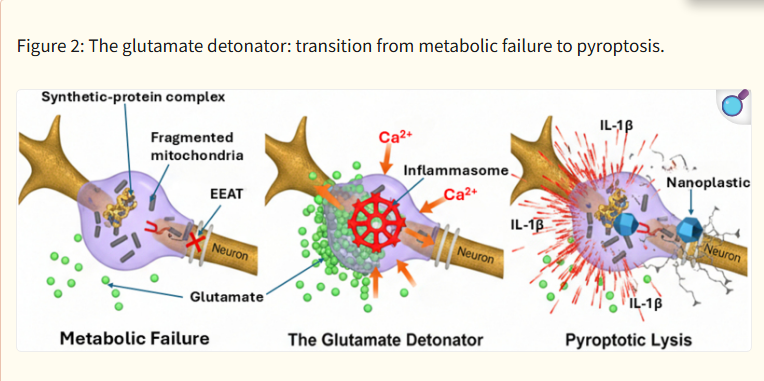
The three-stage cellular cascade leading to neurotoxic liberation (from left to right). Metabolic failure: accumulation of the synthetic-protein complex disrupts mitochondrial integrity (fragmented dark structures), leading to ATP depletion and the failure of excitatory amino acid transporters (indicated by red “X”). The glutamate detonator: extracellular glutamate (green spheres) pools at the synaptic interface, triggering a massive calcium (Ca²⁺) influx into the microglial cell. This secondary signal initiates the assembly of the NLRP3 inflammasome (red crystalline wheel). Pyroptotic lysis: the microglial cell undergoes inflammatory programmed cell death (pyroptosis), resulting in membrane rupture and the liberation of the intact, toxic nanoplastic core into the neuropil, accompanied by a surge of pro-inflammatory cytokines (IL-1β).
 Posted in