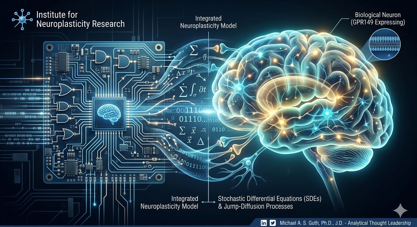
𝗪𝗵𝘆 𝘁𝗵𝗲 𝗙𝘂𝘁𝘂𝗿𝗲 𝗼𝗳 𝗕𝗶𝗼𝘁𝗲𝗰𝗵 𝗗𝗲𝗽𝗲𝗻𝗱𝘀 𝗼𝗻 𝗕𝗿𝗶𝗱𝗴𝗶𝗻𝗴 “𝗪𝗲𝘁𝘄𝗮𝗿𝗲” 𝗮𝗻𝗱 𝗦𝗼𝗳𝘁𝘄𝗮𝗿𝗲.𝗧𝗵𝗲 𝗡𝗲𝘅𝘁 𝗟𝗲𝗮𝗽 𝗶𝗻 𝗔𝗜: 𝗚𝗿𝗼𝘂𝗻𝗱𝗶𝗻𝗴 𝗠𝗮𝗰𝗵𝗶𝗻𝗲 𝗟𝗲𝗮𝗿𝗻𝗶𝗻𝗴 𝗶𝗻 𝗕𝗶𝗼𝗹𝗼𝗴𝗶𝗰𝗮𝗹 𝗣𝗵𝘆𝘀𝗶𝗰𝘀. 𝗙𝗿𝗼𝗺 𝗢𝗯𝘀𝗲𝗿𝘃𝗮𝘁𝗶𝗼𝗻 𝘁𝗼 𝗖𝗼𝗻𝘁𝗿𝗼𝗹: 𝗧𝗵𝗲 𝗡𝗲𝘄 𝗘𝗿𝗮 𝗼𝗳 𝗖𝗼𝗺𝗽𝘂𝘁𝗮𝘁𝗶𝗼𝗻𝗮𝗹 𝗡𝗲𝘂𝗿𝗼𝘀𝗰𝗶𝗲𝗻𝗰𝗲. The future of AI and biotech isn’t just about collecting more data; it’s about building better models of the underlying “physics” of the system. We are seeing a significant shift where classical differential equations are converging with modern machine learning. This hybrid approach is set to redefine how we process neural signals and design cognitive interventions.
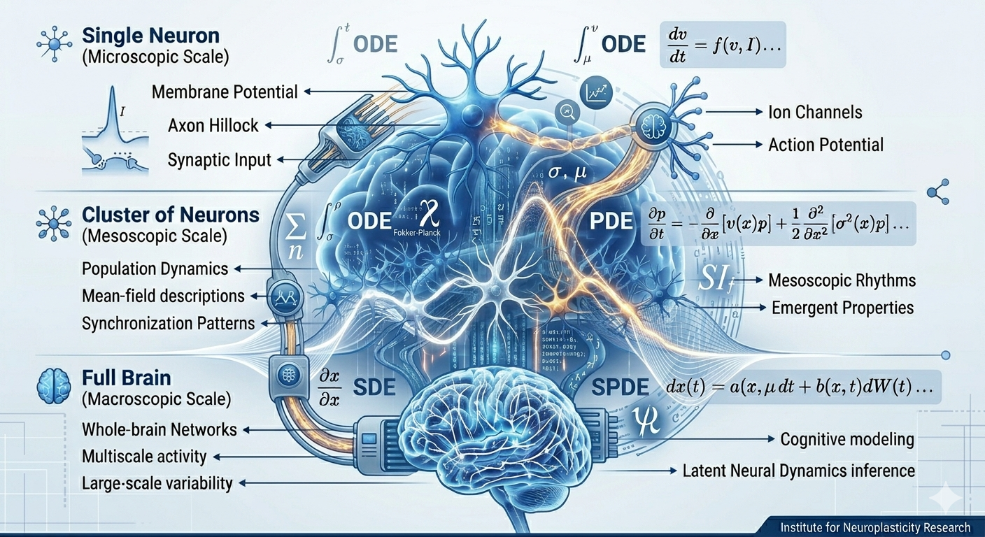
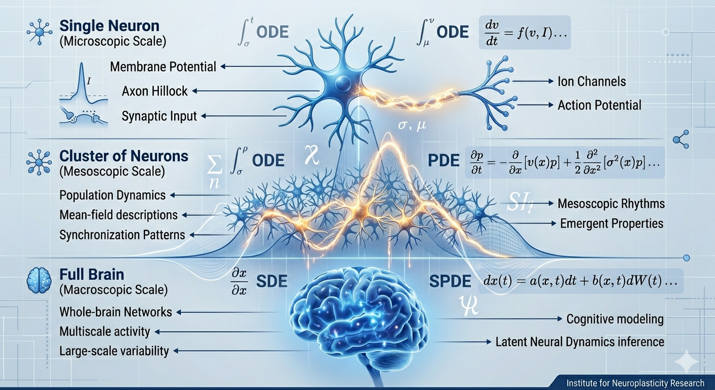
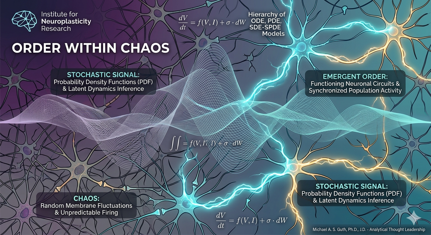
Currently, the hierarchy of Ordinary Ddifferential Equations (ODE), Partial DE, and Stochastic DE models allows us to map everything from deterministic whole-brain networks to stochastic membrane fluctuations. This multiscale approach is vital because it ensures that our models remain grounded in biological reality while benefiting from the computational power of ML-driven inference.
One of the most exciting “open challenges” in this field is the move toward control-oriented formulations. Once we can accurately model neural dynamics using these mathematical frameworks, we can begin to design systems that don’t just observe the brain but interact with it in real-time to correct pathological states or enhance performance.
This convergence has massive implications for the “Global Economy” of health and technology. By integrating kinetic variables and mean-field equations with neural field theory, we are creating a standardized language for computational neuroscience that can be scaled across research and industrial applications.
I am particularly focused on how these models will tackle multiscale inference in the coming years. As we refine our numerical and computational approaches for stochastic systems, the gap between “wetware” (the brain) and “software” (AI) will continue to shrink. The math may be complex, but the potential for innovation is limitless.